
Breaking
Latest health news from South Africa and around the world•Wellness, nutrition, and fitness updates daily•Medical breakthroughs and healthcare updates•Science-backed health explainers and guides•Health tech, reviews, and upcoming events•
 SA Health News Desk
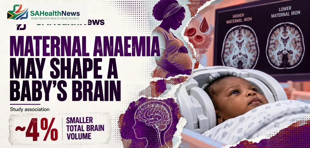
A Cape Town birth-cohort study found ~4% smaller total brain volume in infants whose mothers had pregnancy anaemia — an association, not proven causation.
A South African birth-cohort study has found measurable differences in brain development among infants whose mothers had anaemia during pregnancy, with the changes becoming detectable from around their first birthday.
Researchers from the University of Cape Town and King’s College London followed 394 mother-infant pairs in Cape Town and used two very different MRI systems to investigate how maternal anaemia was associated with infant brain structure.
The peer-reviewed study was published in Brain Communications.
The central finding needs careful wording.
The researchers found an association between anaemia during pregnancy and smaller infant brain volumes. The study was observational, so it cannot establish that maternal anaemia directly caused the differences.
But the pattern was sufficiently consistent to warrant attention.
Babies born to mothers who had been anaemic during pregnancy had, on average, approximately 4% smaller total brain volume than babies whose mothers were not anaemic.
The researchers found differences in several structures involved in learning, movement, executive function, emotion regulation and communication between the two halves of the brain.
UCT reports reductions of approximately 6% in the corpus callosum, 4% in the caudate nucleus and 3% in the putamen among anaemia-exposed infants.
The differences did not all appear immediately after birth.
They became detectable at roughly 12 months.
King’s College London reports that the corpus-callosum difference was around 4% at 12 months and had widened to about 6% by 24 months.
That timing is one of the study’s more important findings.
It suggests that the developmental consequences associated with the prenatal environment may become clearer as the infant brain continues to grow.
Perhaps the most clinically interesting result is that researchers observed the differences even though most anaemia in the cohort was classified as mild.
Anaemia occurs when the blood does not contain enough haemoglobin to carry oxygen effectively.
Iron deficiency is a common cause during pregnancy, although infections, inflammation, nutritional deficiencies and other medical conditions can also contribute.
UCT notes that more than a third of women globally are affected by anaemia, with a disproportionate burden in lower- and middle-income regions including sub-Saharan Africa.
The study also found that mothers living with HIV had a greater risk of being anaemic during pregnancy. Researchers suggest inflammation may affect how the body processes iron, although that mechanism was not the primary outcome tested in this study.
The finding therefore should not be reduced to “pregnant women need more iron”.
Anaemia has multiple causes.
Iron supplementation is appropriate in many situations, but treatment should follow proper antenatal assessment rather than unsupervised high-dose supplementation.
Smaller volume in a brain region is not the same thing as a diagnosis of developmental disability.
The researchers have previously reported similar regional differences in older South African children, and the affected areas have recognised roles in processing speed, executive function, emotional regulation and spatial reasoning.
That makes longer-term neurodevelopment a legitimate concern.
It does not allow researchers to predict that an individual baby with a smaller measured brain volume will develop cognitive impairment.
The new study primarily measured brain structure.
Long-term functional outcomes require separate assessment.
That distinction matters because imaging findings can sound more deterministic than they actually are.
The research also tested technology that could make this kind of science considerably easier to perform in lower-resource settings.
Researchers scanned infants using both a conventional 3-tesla MRI and a portable 64-millitesla ultra-low-field MRI.
The portable system costs less, consumes less energy and requires substantially less infrastructure than a conventional hospital MRI. It is also quieter, allowing researchers to scan sleeping babies without sedation.
The ultra-low-field scanner was sufficiently sensitive to detect brain-volume patterns associated with maternal anaemia.
That finding matters beyond this particular study.
Many of the populations with the highest maternal and child-health burdens live in places with the least access to conventional neuroimaging.
If portable MRI can reliably measure developmental changes, researchers can conduct studies closer to those communities rather than building evidence almost exclusively from children who can reach sophisticated academic hospitals.
The research strengthens the case for preventing, identifying and appropriately treating maternal anaemia.
It does not establish the optimal intervention.
Future trials would need to determine whether preventing or correcting anaemia at specific stages of pregnancy changes infant brain-development outcomes.
Researchers also need to separate the effects of iron deficiency from other causes of anaemia and from overlapping factors such as maternal nutrition, infection, socioeconomic conditions and HIV.
That is why the observational design matters.
The study identifies a potentially important risk marker and shows when measurable brain differences emerge.
It does not close the causal chain.
For South African antenatal care, the practical message is still significant.
Maternal anaemia should not be viewed only as a laboratory number affecting the mother.
The growing body of local research suggests it may also be associated with how the child’s brain develops after birth.
Source: SA Health News